Tracing the Neural Circuitry of Appetite

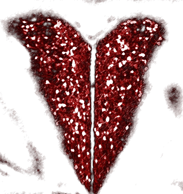
Caption: A stylized image of the MC4R-expressing neurons (in red) within the brain’s PVH, which is the “heart of hunger”
Credit: Michael Krashes, NIDDK, NIH
Credit: Michael Krashes, NIDDK, NIH
If you’ve ever skipped meals for a whole day or gone on a strict, low-calorie diet, you know just how powerful the feeling of hunger can be. Your stomach may growl and rumble, but, ultimately, it’s your brain that signals when to start eating—and when to stop. So, learning more about the brain’s complex role in controlling appetite is crucial to efforts to develop better ways of helping the millions of Americans afflicted with obesity [1].
Thanks to recent technological advances that make it possible to study the brain’s complex circuitry in real-time, a team of NIH-funded researchers recently made some important progress in understanding the neural basis for appetite. In a study published in the journal Nature Neuroscience, the researchers used a variety of innovative techniques to control activity in the brains of living mice, and identified one particular circuit that appears to switch hunger off and on [2].
The circuit involves a group of neurons expressing a protein on their surface called the melanocortin-4 receptor (MC4R). What’s interesting is that the gene that codes for MC4R has been known for years to play an important role in obesity. Mutations in MC4R are rare, but can cause inherited obesity in humans. Also, mice that lack functional versions of the gene overeat and grow extremely obese, ballooning to double their normal weight.
Last year, Michael Krashes, now with NIH’s National Institute of Diabetes and Digestive and Kidney Diseases (NIDDK); Bradford Lowell of Harvard Medical School and Beth Israel Deaconess Medical Center, Boston; and their colleagues discovered MC4R-expressing neurons in the paraventricular nucleus of the hypothalamus (PVH), a very specific region of the brain known for its role in energy balance and hunger [3]. Joined by Alastair Garfield of Scotland’s University of Edinburgh, the researchers have now expanded upon that work, using sophisticated optogenetic and chemogenetic tools to chart in unprecedented detail the neural circuitry that regulates appetite in a mammalian brain.
By selectively activating MC4R neurons in genetically engineered mice with either light or chemicals, the researchers found the animals lost their appetite when MC4R neurons were switched on (which happens to be the normal, default state). Only when MC4R neurons were switched off, did the mice express any interest in eating. So, in the real world, what prompts a mouse to eat? The researchers say the MC4R circuit is just one part of a very complicated neural network, and, when the animal’s body needs more calories, other types of neurons send a signal that turns the MC4R neurons off.
While further study is needed to reproduce this work in both animals and people, the findings should come as welcome news for those seeking to develop drugs to encourage weight loss through appetite suppression. In fact, drugs that turn on MC4R receptors have been considered a promising anti-obesity strategy for years.
The challenge, the researchers explain, is that the receptors also play other vital roles in the body, including the regulation of blood pressure and heart rate. Those effects have stood in the way of pharmaceutical companies’ attempts to produce a successful MC4R-targeted, anti-obesity drug. Perhaps that could change if these new, more-detailed pictures of brain circuitry can point the way to drugs targeted only at the MC4R-expressing neurons involved in appetite.
The findings also serve as an example of the many rewards sure to come as advances in neuroscience—fueled by the NIH-led BRAIN Initiative—move us toward a much more thorough understanding of the brain and its complex role in health and disease.
References:
[1] Overweight and obesity statistics. National Institute of Diabetes and Digestive and Kidney Diseases (NIDDK). October 2012.
[2] A neural basis for melanocortin-4 receptor-regulated appetite. Garfield, AS, Li, C, Madara, JC, Shah, BP, Webber, E, Steger, JS, Campbell, JN, Gavrilova, O, Lee, CE, Olson, DP, Elmquist, JK, Tannous, BA, Krashes, MJ, Lowell, BB. Nature Neuroscience. Published online ahead of print. 2015 April 27.
[3] MC4R-expressing glutamatergic neurons in the paraventricular hypothalamus regulate feeding and are synaptically connected to the parabrachial nucleus. Shah BP, Vong L, Olson DP, Koda S, Krashes MJ, Ye C, Yang Z, Fuller PM, Elmquist JK, Lowell BB. Proc Natl Acad Sci U S A. 2014 Sep 9;111(36):13193-8.
Links:
What are overweight and obesity? (National Heart, Lung, and Blood Institute/NIH)
Krashes Lab (NIDDK/NIH)
Lowell Lab (Beth Israel Deaconess Medical Center and Harvard Medical School, Boston)
The Brain Initiative (NIH)
NIH Support: National Institute of Diabetes and Digestive and Kidney Diseases


.png)


No hay comentarios:
Publicar un comentario